Case study: Thermography used to identify inflammation from a harness injury
Thermal imaging was used to identify areas of inflammation on a dog who presented with hind limb paralysis. Significant inflammation was found in soft tissue and bony structures directly impacted by a poorly designed and fitted harness.
Partnering with the Canine Holistic Wellness Centre in Sydney, we conducted Thermal Imaging of dogs before and after hyperbaric treatment. What we found through Thermal Imaging was incredible.
The attached images are of a 2.5year toy breed dog who initially presented with hind limb paralysis, including faecal and urinary incontinence. She has been undergoing hyperbaric treatment for 1+ weeks, regaining more movement with each treatment.
The treatment's effects were amazing, with a significant reduction in ataxia and increased mobility directly after treatment. However, to get the best care, we needed a clearer picture of what was happening, the cause of the sudden onset of paralysis was still unknown.
Using the thermal imaging camera, I took a series of images that gave this patient a voice. Although not diagnostic, thermal imaging has shown us exact areas of concern, and from these images, we isolated the potential cause.
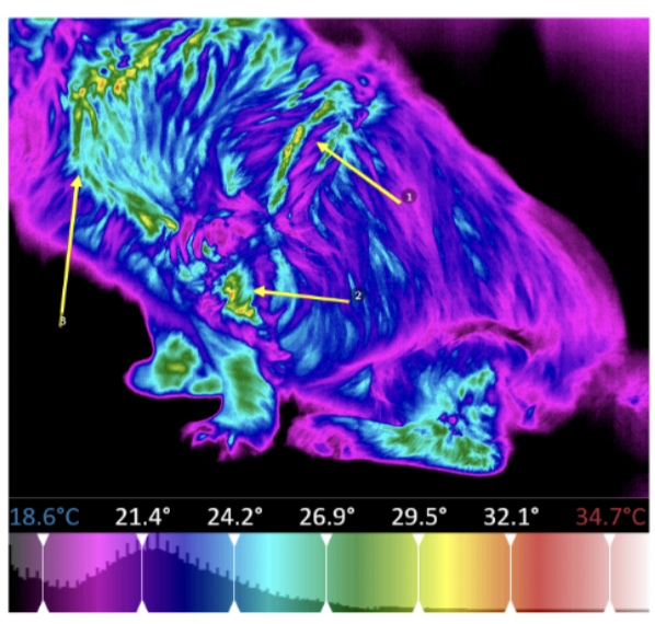
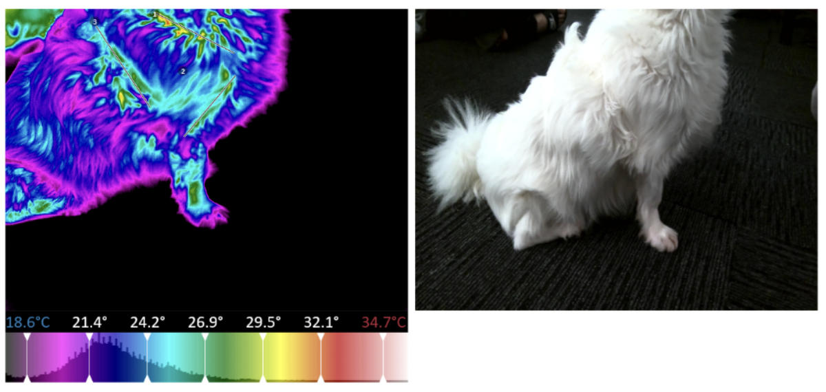
All the symptoms of this young dog were in the posterior region. Yet, these non-invasive, non-contact images show the patient is experiencing inflammation in the anterior thoracic region. This area correlates with the harness used on this dog – a popular brand. I will discuss harnesses in a separate post.
The images showed us a small area of hyperthermia on the LHS knee – only when the patient was actively using this joint to squat. Otherwise, the posterior region of this patient did not display any significant findings.
Anterior region images identified several areas of concern. The hyperthermia associated with the harness strap around the dog's barrel was surprising. Having not worn this harness for over a week, its impacts are still clearly visible in the infrared heat map.
Hyperthermic areas of concern include the LHS brachiocephalic muscles, bilateral latissimus dorsi, trapezius and rhombus muscles – all consistent with the harness used on this patient. Thermal imaging has identified the T1-T4 region as the most significant area; this is where the hard plastic buckle sits and directly applies pressure.
The RHS scapular and humorous were also found to be hyperthermic; we can see in the images that the inflammation directly follows the spine of the scapular to the point of the shoulder and then directly follows the bone of the humorous. Compared to the LHS, we have a significant finding that was easily missed without thermal imaging.
Thermal imaging used as a pre-diagnostic tool can help determine where x-rays are needed and where treatments need to be directed. In cases like this, our Wellness Checks can highlight the dangers and damage of incorrectly fitting or poorly designed equipment.
If you haven’t already contacted us to book a wellness check, click on the button at the top and send us a message.
If you'd like to learn more about chosing a harness check out of article on design and fitment of harnesses for dogs here.


Leave a comment